What is Bronchoscopy? How is it done?
Tuesday, August 1, 2017 | Healthy lifestyle
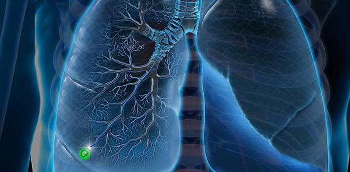
Bronchoscopy with a general definition is the visualization of the bronchial tree of the airways from the inside. During this procedure, the anatomy of the bronchial tree is examined and many diseases, especially lung cancer, can be diagnosed. Bronchoscopes commonly used today are flexible and the part of the patient that enters the bronchial tree is very incarcerated and does not make the patient feel uncomfortable. The image taken from the lens and the airplane at the end of the device is viewed from the monitor. During the procedure, biopsies can be taken from the bronchial mucosa, obtained by brushing, or the presence of bacterial or tumor cells in this fluid obtained by physiological saline delivery to the bronchial tree. In recent years, it has become possible to remove these strictures with laser or cortical applications in tumor or non-tumor diseases that constrict airways during bronchoscopy. In addition, bronchoscopies and light-emitting tumors in different wave lengths developed in recent years can be detected at a very early stage when they can not be seen with normal bronchoscopes or can not be recognized.
To whom Bronchoscopy should be done?
Bronchoscopy used for diagnosis and treatment;
- Chest X-ray or computed tomography taken for various reasons suggests lung tumor, presence of lesion,
- If there is any lesion on chest X-ray and diagnosis is not possible with other methods,
- In the presence of a voice annoyance that lasts for 2 weeks and is not considered a disease by the otolaryngologist directly by the ENT specialist,
- In the presence of ongoing chronic cough that has been investigated with lung graphy, tomography, allergy tests, pulmonary function tests, but no results have been reached,
- In the event of spitting of blood sputum or blood together with coughing,
- In cases in which a diagnosis of breathlessness can not be reached in patients with complaints of shortness of breath,
- During breathing, especially when there is a sensation of wheezing in a particular lung area and no other reason to explain it,
- In chest trauma and injuries,
- In the presence of excessive secretion (excretion) accumulation in the bronchi,
- In order to remove foreign objects from air carriers,
- In order to remove invasive bronchoscopy applications, ie laser, argon plasma cautery, electrocautery, cryocoter, from benign or malignant tumors originating from the main bronchus and causing excessive breathlessness and drowning in the patient,
- In order to apply stents for the treatment of stenosis of the main bronchi due to various reasons, it is decided by the specialist physician.
Preparing the patient before bronchoscopy
The purpose of the bronchoscopy to be done to the patient before the procedure is explained in detail, how the possible losses and the procedure will be applied if not done. Before fiberoptic bronchoscopy performed with local and general anesthesia, it is suggested that the patient should be hungry for at least 6 hours. For this purpose, if the operation is done in the morning, eating and drinking will be stopped from 24:00 at night.
Bronchoscopy with local anesthesia usually does not require operating room conditions and is used as an office procedure. The patient is taken to the bronchoscopy room and once more detailed information is given about the operation to be performed and the application is started. General anesthesia and bronchoscopy are performed in advanced endoscopy units or operating room conditions. For patient safety, it is preferable to have an anesthesiologist in the operating room, whether under local anesthesia and sedation, or under general anesthesia.
How is fiberoptic flexible bronchoscopy performed with local anesthesia?
The patient's mouth and throat taken to the bronchoscopy unit is numbed with a spray. The aim of local anesthesia is to remove the cough and reflex reflex from the patient's nose. There is no pain during bronchoscopy. Anesthesia made for this reason is not for pain. After the mouth and throat are numbed, another needle is inserted through the vein to provide sedation (sedation), followed immediately by the bronchoscope through the larynx before the mouth or mouth and from the soundtrack through the main airway (trachea). At this time, the movements and anatomy of the patient's vocal tracts are also examined. The bronchoscope may cough from time to time during initial entry into the airway and during administration, except that the patient does not feel pain or pain, as noted above. Fiberoptic bronchoscopy trachea, right and left main bronchi, 2 lobes on the left, 3 bronchial lobes on the right and more extreme points of these lob bronchi can be seen. From these areas or from the more distal regions of the lungs can be biopsied, lavaged, and delivered to the bacteriological and cytopathologic examination. If necessary, the procedure is improved by using endobronchial ultrasonography or radioscopy guidance during the procedure. Flexible bronchoscopy procedure takes 10-12 minutes on average. The patient can be sent home after the procedure. After the procedure, the patient has to eat and drink for 2 hours after the mouth and throat are in a drowsy state.


